Diagram Of Liver Cell - Cancers | Free Full-Text | Liver Cancer: Current and ... : These functions make the liver a vital organ without which the tissues of the body would quickly die from lack of energy and nutrients.
byAdmin•
0
Diagram Of Liver Cell - Cancers | Free Full-Text | Liver Cancer: Current and ... : These functions make the liver a vital organ without which the tissues of the body would quickly die from lack of energy and nutrients.. It is a large organ, with its major lobe occupying the right side of the abdomen below the diaphragm, while the narrower left lobe extends all the way across the abdomen to the left. Learn vocabulary, terms and more with flashcards, games and other study tools. The liver has many functions. Liver cells express mscca (bear, 1990) and previous studies had shown that osmotic swelling of epithelial cells activates an mscca‐dependent figure 5.7. 12.08.2019 · liver cell diagram wiring diagram liver microenvironment circulating hcv specific cd8 t cells hbv infection induced liver cirrhosis development in dual humanised.
Liver cells express mscca (bear, 1990) and previous studies had shown that osmotic swelling of epithelial cells activates an mscca‐dependent figure 5.7. These functions make the liver a vital organ without which the tissues of the body would quickly die from lack of energy and nutrients. It performs 500 essential tasks, including detoxification, protein synthesis, and the classed as part of the digestive system, the roles of the liver include detoxification, protein synthesis, and the production of chemicals that help digest food. Learn vocabulary, terms and more with flashcards, games and other study tools. Control of liver cell fate decision by a gradient of tgf beta signaling modulated by onecut transcription factors.
3D Diagram Of A Plant Cell (With images) | Animal cell ... from i.pinimg.com Human anatomy detailed diagram of various human organs liver, heart, kidneys, lungs, colon, intestine, stomach, brains, etc can be used in. The liver is a vital organ found in humans and other vertebrates. The liver is partially surrounded by the ribs, and extends from the level of the fifth intercostal space to the lower margin of the right rib cage, which protects this highly vascular organ. Hepatocyte nuclei often contain a prominent nucleolus. Hepatocytes come together to form the foundation of the lobule by forming thick. On the other hand, eukaryotes have chromosomes that are made up of dna and protein. Learn about the human liver. Lifestyle changes may slow the progression of some types of liver disease.
Ƽ intricately involved in carbohydrate, fat, and protein metabolism.
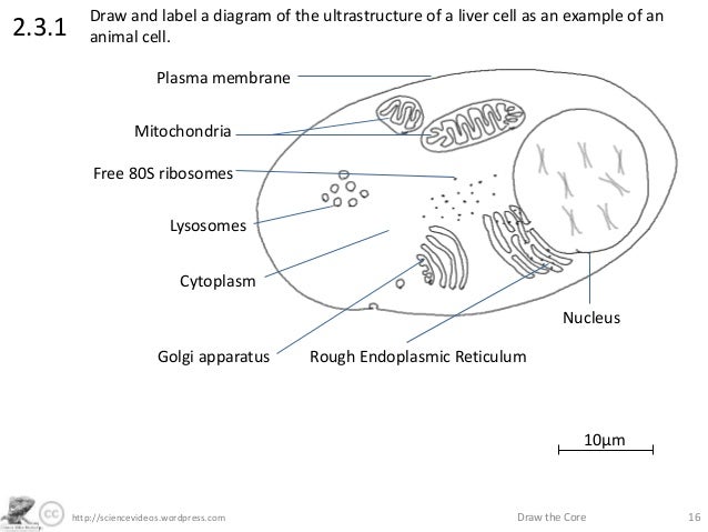
Learn about the human liver. It should be large, clear and with specific labels. Medical labeled diagram with all kind cells. Animal liver cell diagram ~ diagram. The liver is the largest solid organ in the human body. Whatever an organism does for survival it does for the survival of its cells. Binucleated hepatocytes (= containing two nuclei). 2.3.1 draw and label a diagram of the ultrastructure of a liver cell as an example of an animal cell. Example of blood, neurons, cardiac, bone, intestinal, epithelial, fat, liver and. Documents similar to liver pathophysiology and schematic diagram. Two larger ones (right and left) and two. Hepatocytes are polygonal epithelial cells with abundant eosinophilic, granular cytoplasm and large, centrally located round nuclei. Causes, treatment, and life expectancy vary.
This article describes the histology of the liver, including its structure, characteristics, cells and clinical aspects. You will be using the microscope in your biology study. Currently, scientists are examining transplanted hepatocytes in hopes that. 2.3.1 draw and label a diagram of the ultrastructure of a liver cell as an example of an animal cell. Expression of liver specific proteins decreases with time in culture, but is reactivated by growing the cells in serum free medium.
http://sciencevideos.wordpress.com Draw the Core 162.3.1Draw from image.slidesharecdn.com Binucleated hepatocytes (= containing two nuclei). The cell lives and, as a result, the organism lives. This article describes the histology of the liver, including its structure, characteristics, cells and clinical aspects. Below is a diagram of a compound light microscope. Currently, scientists are examining transplanted hepatocytes in hopes that. Ƽ store vitamins and minerals; 2.3.2 annotate the diagram from 2.3.1 with the functions of each named structure. 12.08.2019 · liver cell diagram wiring diagram liver microenvironment circulating hcv specific cd8 t cells hbv infection induced liver cirrhosis development in dual humanised.
It performs 500 essential tasks, including detoxification, protein synthesis, and the classed as part of the digestive system, the roles of the liver include detoxification, protein synthesis, and the production of chemicals that help digest food.
Lifestyle changes may slow the progression of some types of liver disease. The liver has many functions. The liver is the largest internal organ of the human body, weighing approximately 1.5 kg. Another type of liver cell is the endothelial cells. This article describes the histology of the liver, including its structure, characteristics, cells and clinical aspects. The stellate fat storing cell. These functions make the liver a vital organ without which the tissues of the body would quickly die from lack of energy and nutrients. Animal liver cell diagram ~ diagram. You will be using the microscope in your biology study. Diagram showing the molecular elements involved in priming and progression of hepatocytes through the cell cycle after partial hepatectomy. Pharmacotoxicological studies and for the investigation of. Example of blood, neurons, cardiac, bone, intestinal, epithelial, fat, liver and. Where is your liver is located.
Liver medicine refers to all diagnostic and treatment strategies of diseases and conditions that cause liver failure directly or indirectly. It is a large organ, with its major lobe occupying the right side of the abdomen below the diaphragm, while the narrower left lobe extends all the way across the abdomen to the left. Hepatocytes are polygonal epithelial cells with abundant eosinophilic, granular cytoplasm and large, centrally located round nuclei. Start studying liver cell model. Expression of liver specific proteins decreases with time in culture, but is reactivated by growing the cells in serum free medium.
Histology - Liver Lobule - lobule and central vein and ... from i.pinimg.com Two diagrams of liver structure removed for copyright reasons. Liver medicine refers to all diagnostic and treatment strategies of diseases and conditions that cause liver failure directly or indirectly. Hepatocytes are polygonal epithelial cells with abundant eosinophilic, granular cytoplasm and large, centrally located round nuclei. Pharmacotoxicological studies and for the investigation of. The cell lives and, as a result, the organism lives. Expression of liver specific proteins decreases with time in culture, but is reactivated by growing the cells in serum free medium. Below is a diagram of a compound light microscope. Blood flows through the liver.
An in vitro model for.
12.08.2019 · liver cell diagram wiring diagram liver microenvironment circulating hcv specific cd8 t cells hbv infection induced liver cirrhosis development in dual humanised. Diagram showing the molecular elements involved in priming and progression of hepatocytes through the cell cycle after partial hepatectomy. The liver is the largest internal organ of the human body, weighing approximately 1.5 kg. Causes, treatment, and life expectancy vary. It is a large organ, with its major lobe occupying the right side of the abdomen below the diaphragm, while the narrower left lobe extends all the way across the abdomen to the left. The liver performs many essential functions related to digestion, metabolism, immunity, and the storage of nutrients within the body. Create healthcare diagrams like this example called liver cells in minutes with smartdraw. Anatomically the liver consists of four lobes: You will be using the microscope in your biology study. Human anatomy detailed diagram of various human organs liver, heart, kidneys, lungs, colon, intestine, stomach, brains, etc can be used in. It should be large, clear and with specific labels. Control of liver cell fate decision by a gradient of tgf beta signaling modulated by onecut transcription factors. | human cell structure, animal cell project, animal cell.
Start studying liver cell model diagram of liver. Example of blood, neurons, cardiac, bone, intestinal, epithelial, fat, liver and.